

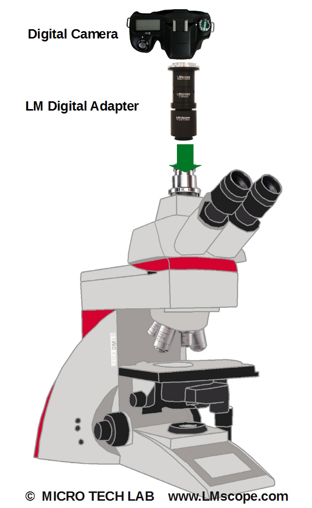
Capture exceptional images with the Leica DM4 B/DM6 B and our LM digital adapters
The upright digital research microscopes DM4 B and DM6 B were developed by Leica for use in biomedical research and in clinical laboratories. The two systems are successors to Leica’s DM4000 B, D5000 B, DM5500 B and DM6000 B models and further refine this predecessor generation of automated research microscopes. Both microscopes are equipped with displays that perform different functions: the display of the DM4 B shows all current microscope settings, while the DM6 B features a touchscreen for intuitive remote control of the microscope.
The DM4 B uses LED transmitted light illumination for the following contrasting methods: bright field, dark field, phase contrast and polarisation contrast. Fluorescence illumination is provided by LED or mercury vapour. The DM6 B offers a choice between LED and halogen transmitted light illumination. In addition to the contrasting methods described above, differential interference contrast (DIC) observation is also supported. The DM6 B comes equipped with a motorised stage and has a mechanical or motorised focus option. The user can choose the objective best suited for the task at hand from the full portfolio of Leica objectives. The objective turret (coded, partially motorised) offers a choice of 6 or 7 objective positions.
The field of view of the camera port measures 19mm and is matched to the chip size of sCMOS cameras. Large-format CMOS sensors have a better signal-to-noise ratio, which is particularly important at high ISO settings. With our LM digital adapters, users are free to either use same-brand cameras or standard DSLR cameras of their choice to produce captivating, high-resolution microscope images. Today’s sophisticated DSLR and system cameras achieve superb colour rendition and an amazing level of detail. The motorised field diaphragm features six round and square field stops of different sizes, making it easier to match the image section to the chip size of the camera being used. The Fluorescence Intensity Manager (FIM) regulates the lighting to protect the specimen against photobleaching.
A distinguishing feature of the DM4 B and DM6 B microscopes is the automation of specific functions. For example, switching between contrast methods is initiated by pressing a single button – all changes in settings are performed automatically. Both instruments are available with the LAS X software package, which guides users through image acquisition step by step and provides a quick overview of the specimens. Programmable function control buttons and touch panel operations simplify all phases of microscope operation.
Of course, ergonomics also play an important role with these high-quality microscopes: ergo tubes and adaptable stages for right- or left-handed operators make working for hours at a time less strenuous.
The microscopes come with a broad range of tools and accessories, too many to comprehensively describe here (such as a fully automated fixed stage microscope for electrophysiological research or sample preparation with laser microdissection). When combined with the appropriate light source, the instruments are also well suited for optogenetics (a relatively new field of technology involving the use of light to control genetically modified cells).
Conclusion:
Leica’s two upright research microscopes DM4 B and DM6 B are exceptionally well suited for photomicrography. With the help of our LM digital adapters, they can be combined with favourably priced DSLR and system cameras quickly and without difficulty.
Photography:
Fitting the microscope to digital single-lens reflex (DSLR), mirrorless interchangeable-lens cameras (MILC ), digital single-lens mirrorless (DSLM) or C-mount cameras is easy with our LM digital SLR adapters, which feature a plan achromatic optical system. Our products make it possible to capture top-quality microscope images. To help you select the adapter that is right for your camera, we have set up an online configurator on our website. You can also email us – ideally with attached photographs of your microscope.
Modern DSLR and single-lens mirrorless (DSLM) offer the latest technology and are generally very well suited for microscopy applications. Most of them can be controlled remotely via PC/Mac. Because of their high sales volumes, they offer an excellent price/performance ratio compared to special-purpose microscope cameras.
Features of top DSLR and single-lens mirrorless cameras (DSLM):
- Large, powerful full-frame sensors (36 x 24 mm)
- Sensor resolution of 61 megapixels or 240 megapixels with Pixel Shift technology
- High light sensitivity (ISO 400,000+)
- Extensive dynamic range (up to 15 aperture stops/f-stops)
- Short exposure times (1/8000 second) up to 1/32,000 seconds using the digital shutter
- 4K Ultra HD or 8K Ultra HD video function
- Live video capture on external monitors in ultra HD quality
In most cases, these cameras are significantly more powerful than microscope cameras with smaller sensors (1/2" or 2/3"). On our website you will find our current camera recommendations and a camera ranking which is specifically tailored to microscopy applications.