


 800
What’s that floating in my backyard pond? The larval skin (moulted shell/exuvia) of a dragonfly under the microscope
The LM photomicroscope and automated focus stacking on a PC or Mac: a more capable alternative to conventional (Greenough-type) stereo microscopes
Our backyard pond is a popular spot for dragonflies. These predators hunt small insects which they catch in midair. Female dragonflies lay their eggs by releasing them over the water while flying, or they embed them in aquatic plants or deposit them along the water’s edge. Once hatched, the larvae live underwater and metamorphose into adult dragonflies Many species moult ten times or more before the dragonfly emerges as an adult – a process that may take weeks or even years to complete. The shed skins left behind at each moult, known as exuviae, make excellent subjects for microscope photography.
With specimens this challenging, conventional microscopes often reach their limits. At around 40 mm in size and with a layer thickness of about 2–4 mm, the specimen cannot even be positioned under the objective lenses of many conventional reflected-light microscopes because there isn’t enough clearance.
Standard stereo microscopes do offer sufficient working distance, even for large specimens, but their dual, offset optical paths and long working distance often come at the expense of the resolving power of the objectives, which is needed to capture even the smallest details with exceptional clarity. Conventional (Greenough-type) stereo microscopes were developed for visual observation through the eyepieces. The two offset optical paths create the perception of three-dimensionality when the image is processed by the brain. However, when taking photographs through one of the two optical paths (left or right), the image quality is often disappointing.
Achieving images with excellent depth of field requires a technique known as focus stacking (focus bracketing): multiple images (around 20–50) are taken at different focus distances and combined into a stack, which is then processed with a special software tool to create one perfect composite image. This workflow requires a motorised focusing rail (z-axis). Many microscopes and stereo microscopes in the lower and mid-tier price ranges are difficult to retrofit with a motorised focusing system.
LM photomicroscopes and LM macroscopes are equipped with high-resolution objectives and have been specifically developed for professional digital photography. They also offer seamless integration with high-performance digital cameras with large sensors (full-frame, APS-C, M43), which can be attached directly with no additional accessories required. Their centric optical path allows for superior image quality. Our devices are factory-ready for installation of a motorised z-axis focusing rail.
The modular design of the LM photomicroscope/macroscope offers versatile configuration options to meet the user’s specific needs, and existing equipment and components from other manufacturers can remain part of the setup. The individual modules, such as stages, tube modules, objectives, filter wheel, filter tray and accessories, are also available separately.
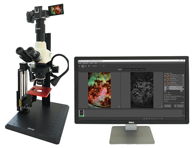
With the appropriate software, both the camera and the stage can be controlled remotely from a computer. This enables the automated creation of large stacks containing more than 100 images captured at precisely stepped focus intervals. After capture, a single click merges the stack into one perfect shot.
Focus stacking achieves exceptional depth of field together with high-resolution detail. The following application example demonstrates the results.
The nearly transparent skin of a dragonfly larva reveals interesting structures that we have made visible with our LM photomicroscope. We placed the specimen in a Petri dish containing about 2–3 mm of water. Skilful lighting brings out anatomical structures that are otherwise invisible to the naked eye. Darkfield illumination creates an impressive three-dimensional appearance. To achieve greater depth of field, we used focus stacking: we captured a stack of about 40 images and processed them with the Helicon Focus image processing software. We also applied polarisation to enhance the colours and eliminate reflections from the water. For image capture, we used a Nikon Z7 mirrorless system camera, which has a high-quality full-frame sensor. With 45 megapixels, the sensor resolution is more than adequate to reveal even the smallest details at low overview magnifications.
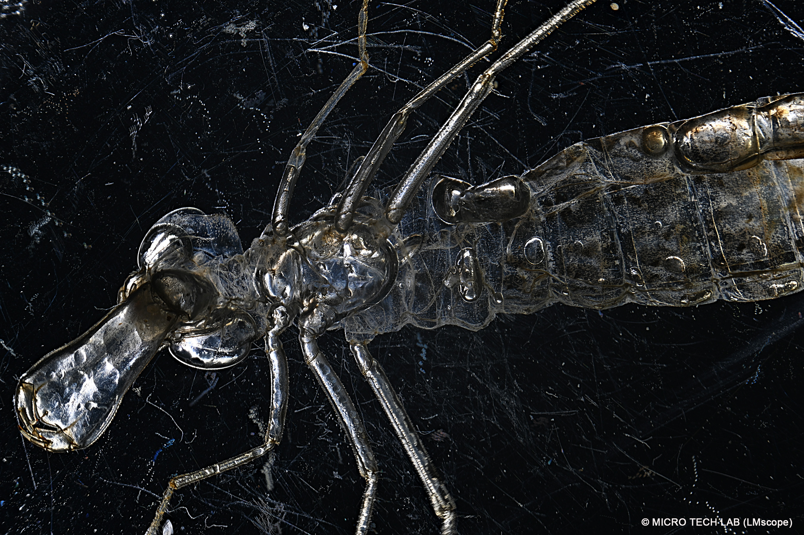
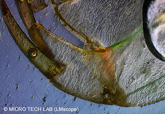
The image below shows a distinctive feature of the dragonfly larva: its so-called catching mask. Formed from an elongated lower lip, it is used to capture prey. When striking at prey, this mechanism, which is folded beneath the head and thorax when at rest, shoots forward at high speed. The paired grasping hooks (labial palps), which hold the prey in place, are distinctly visible.
Imaging setup: stack of 40 individual images captured using the LM photomicroscope at 50x magnification, polarisation, a StackShot focusing rail, the Helicon Focus software and a Nikon Z7 camera
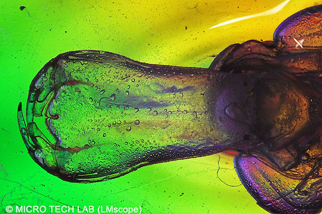

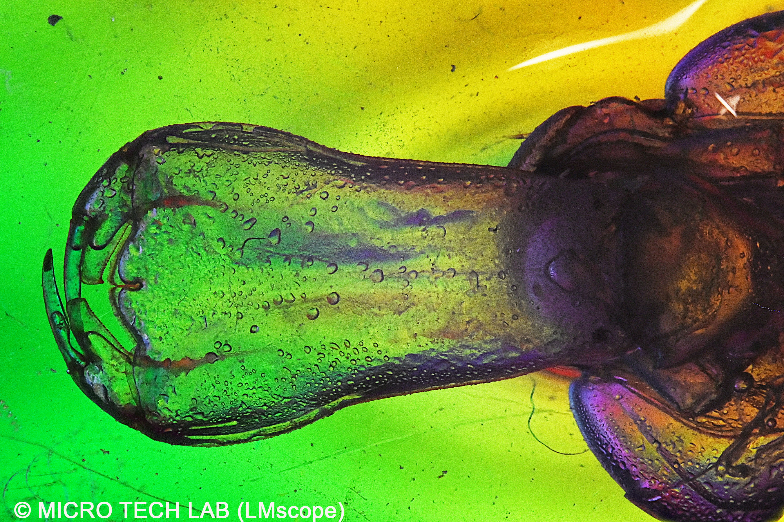
View the image in higher resolution
Below is a close-up of the catching mask at 100x magnification, showing the grasping hook and its sharply pointed tooth.
Imaging setup: stack of 40 individual images captured using the LM photomicroscope at 100x magnification, darkfield illumination, a StackShot focusing rail, the Helicon Focus software and a Nikon Z7 camera
View the image in higher resolution
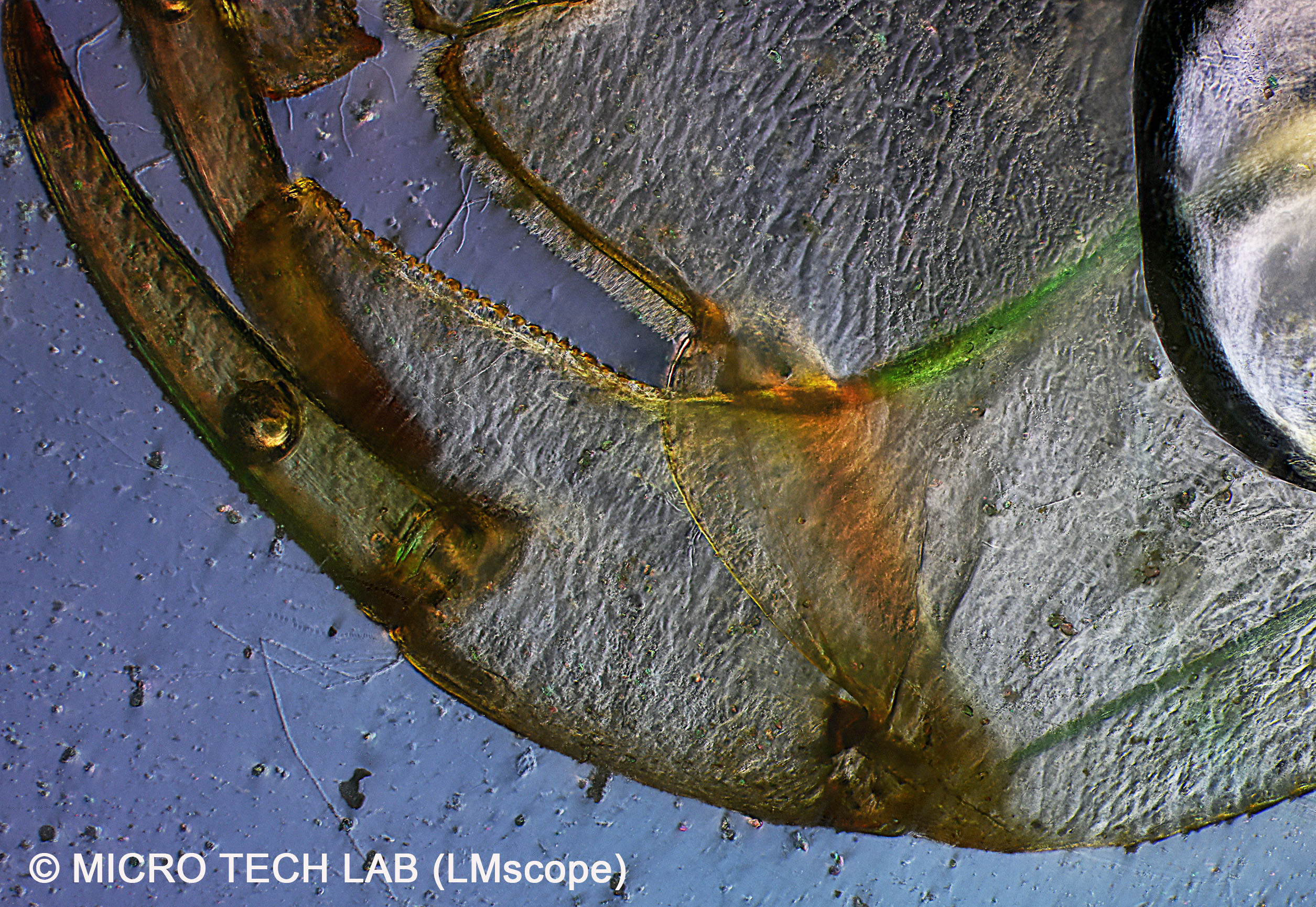
Like the adult dragonfly, the larva already has compound eyes, although they are considerably smaller than those of the fully grown dragonfly (imago). These eyes are made up of individual facets, and their number increases with every moult. They are even visible on the larval skin, as shown in the image below.
Imaging setup: stack of 40 individual images captured using the LM photomicroscope at 100x magnification, polarisation, a StackShot focusing rail, the Helicon Focus software and a Nikon Z7 camera
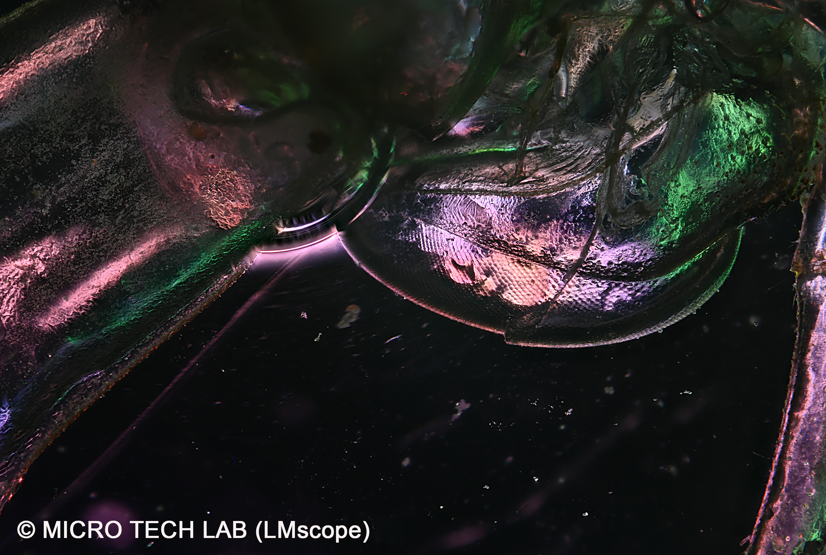
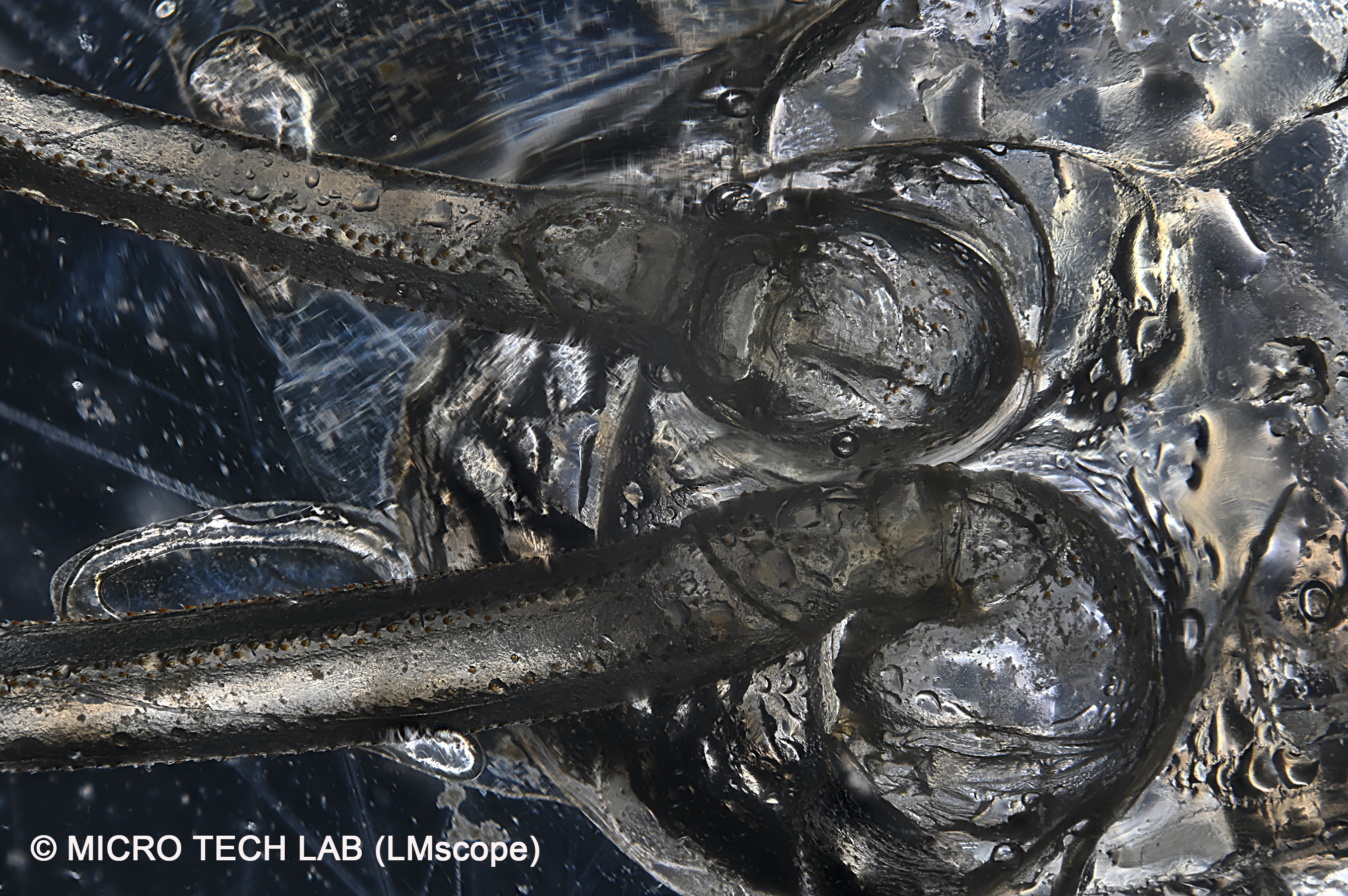
The next image shows one side of the larva’s three pairs of spiny legs, which are used to capture and hold prey but, most importantly, provide a firm grip when the larval skin is shed during metamorphosis.
Imaging setup: stack of 40 individual images captured using the LM photomicroscope at 40x magnification, polarisation, a StackShot focusing rail, the Helicon Focus software and a Nikon Z7 camera
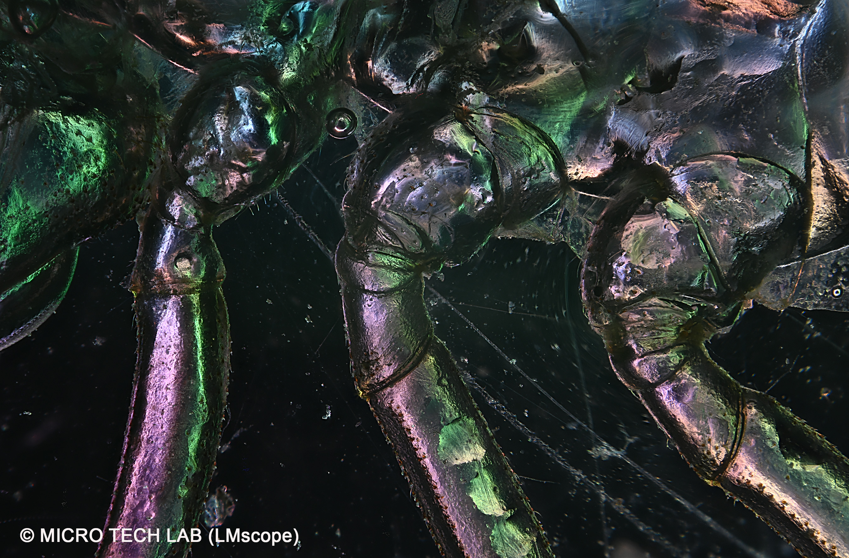
For this image, we turned the specimen over: we can now see where two of the three legs on one side connect to the thorax.
Imaging setup: stack of 40 individual images captured using the LM photomicroscope at 100x magnification, darkfield illumination, a StackShot focusing rail, the Helicon Focus software and a Nikon Z7 camera.
If you have any questions, please do not hesitate to reach out. We will be happy to assist you.
12.10.2026New LM Digital Adapter for: Sony Alpha 9 III / Sony Alpha 7R VI / Nikon Z9 / Nikon Z8 / Sony Alpha 7R V / Sony Alpha 1 II / Sony Alpha 1 / Sony Alpha 9 II (ILCE-9M2) / Sony FX3 Cinema Line / Sony Alpha 9 / Nikon D6 / Canon EOS R3 / Sony FX3 / Canon EOS R6 Mark II / Canon EOS R8 / Sony Alpha 7R IV / Canon EOS R5 II / Nikon Z6III / Canon EOS R5 / Sony Alpha 7S II / Sony Alpha 7S III / Sony Alpha 7R III / Canon EOS R6 / Nikon Z6 / Nikon Z6II / Sony Alpha 7R II / Nikon Z7 / Nikon Z7II / Canon EOS R / Canon EOS Ra (Astro) / Nikon Z5 / Sony Alpha 7C / Canon EOS RP / Sony Alpha 7S / Canon EOS R7 / Leica SL2-S / Canon EOS R10 / Nikon Z50 II / Canon EOS 1D X Mark III / Nikon Z50 / Nikon Z30 / Nikon Z fc / Nikon D850 / Canon EOS 1D X Mark II / Nikon D780 / Olympus OM-1 / Sony Alpha 7III / Olympus OM-D E-M1 Mark III / Canon EOS R100 / Sony Alpha 6700 / Nikon D5 / Sony Alpha 6600 / Fujifilm X-H2S / Fujifilm X-S10 / Fujifilm X-E4 / Fujifilm X-Pro3 / Olympus OM-D E-M1X / Sony Alpha 6400 / Sony Alpha 6100 / Sony ZV-E10 / Canon EOS 1D X / Nikon D4s / Olympus OM-D E-M5 III / Canon EOS 90D / Canon EOS 5D Mark IV / Nikon D4 / Nikon D750 / Canon EOS 6D Mark II / Fujifilm X-T5 / Fujifilm X-T4 / Fujifilm X-T3 / Sony Alpha 6300 / Sony Alpha 6500 / Nikon D500 / Nikon D810 / Nikon D800 / Canon EOS M6 Mark II / Nikon D800E / Nikon Df / Panasonic Lumix DC-G9 / Nikon D610 / Nikon D600 / Canon EOS 250D / Canon EOS 850D / Rebel T8i / Canon EOS 6D / Sony Alpha 99 II (SLT-A99 II) / Canon EOS M200 / Canon EOS 5DS R ( without low-pass filter) / Olympus OM-D E-M1 Mark II / Canon EOS 80D / Canon EOS M50 Mark II / Canon EOS 5DS / Canon EOS M50 / Sony Alpha 77 II / Canon EOS 70D / Nikon D7200 / Pentax K-1 Mark II / Canon EOS 200D / Canon EOS 800D / Rebel T7i / Canon EOS 77D / Canon EOS 5D Mark III /