Strawberry: close-up cross section transmitted light
Strawberry: close-up cross section transmitted light
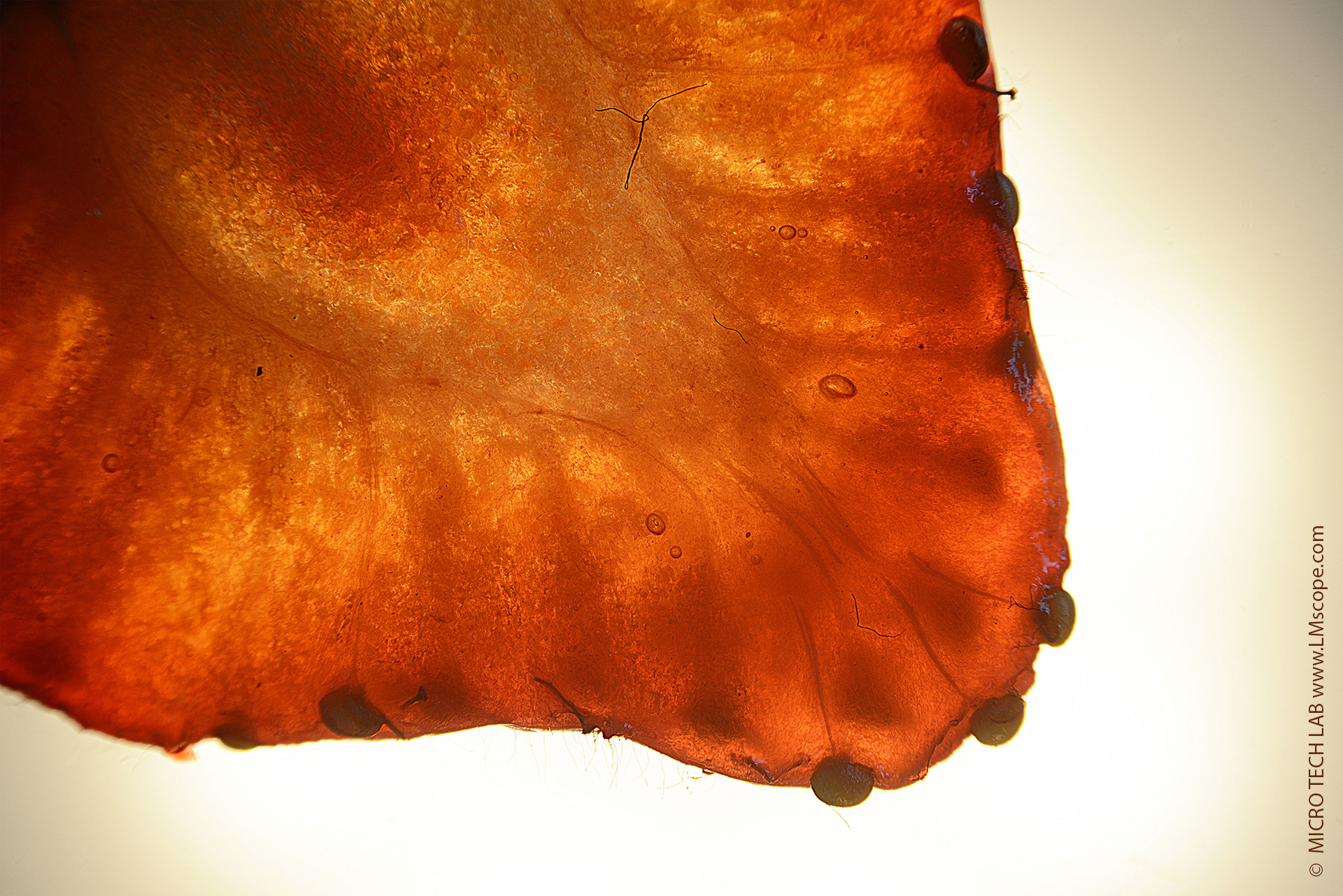
Microscopic image of the cross-section of a garden strawberry with LM photomicroscope and Nikon Z7II: approx. 1–2 mm thickness, 20x microscope magnification
In botanical terms, the red “berry” we enjoy is actually the enlarged base of the flower stalk (receptacle) with numerous seeds embedded in its surface. The hard, yellowish specks on the surface, which resemble seeds, are in fact the real fruits of the strawberry, scientifically known as achenes. These are clearly visible in the image. The wall of each achene houses a true seed (ovule) with the potential to sprout into a new strawberry plant (seedling).
Fibrovascular bundles, composed of longer, tougher cells than the fleshy part, connect the central cylinder of the fruit to the seeds, holding the fruit together. With a combination of transmitted and reflected light illumination (LED spots with gooseneck and diffuser, Kaiser transmitted light unit), these bundles can be made clearly visible.